Healthcare
Blockchain in Healthcare for Achieving Patients' Privacy
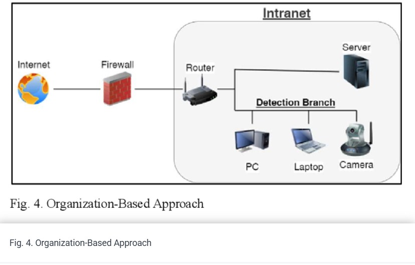
Heath data are sensitive and valuable for individuals. The patients need to integrate and manage their medical data continuously. Personal Health Record (PHR) is introduced as a solution for managing their health information. It gives patients ownership over their medical data and provides physicians with realignment data. However, it does not achieve reliability, traceability, trust, nor security of patient control. Centralization of any data is vulnerable to the problem of hacking and single failure in addition to control from one organization. So, the centralization of data is the common

Comparative Analysis of a Generalized Heart Localization Model: Assessing Its Efficacy Against Specialized Models
Heart localization holds significant importance in the process of the diagnosis and treatment of heart diseases. Additionally, it plays an important role in planning the cardiac scanning protocol. This research focuses on heart localization by employing the multi-label classification task with the utilization of RES-Net50. The primary objective is to predict the slices containing the heart and determine its endpoint. To ensure high-quality data, we implement filtering techniques and perform up-sampling during the pre-processing stage. Two experiments were conducted to assess different

Rice Plant Disease Detection and Diagnosis Using Deep Convolutional Neural Networks and Multispectral Imaging
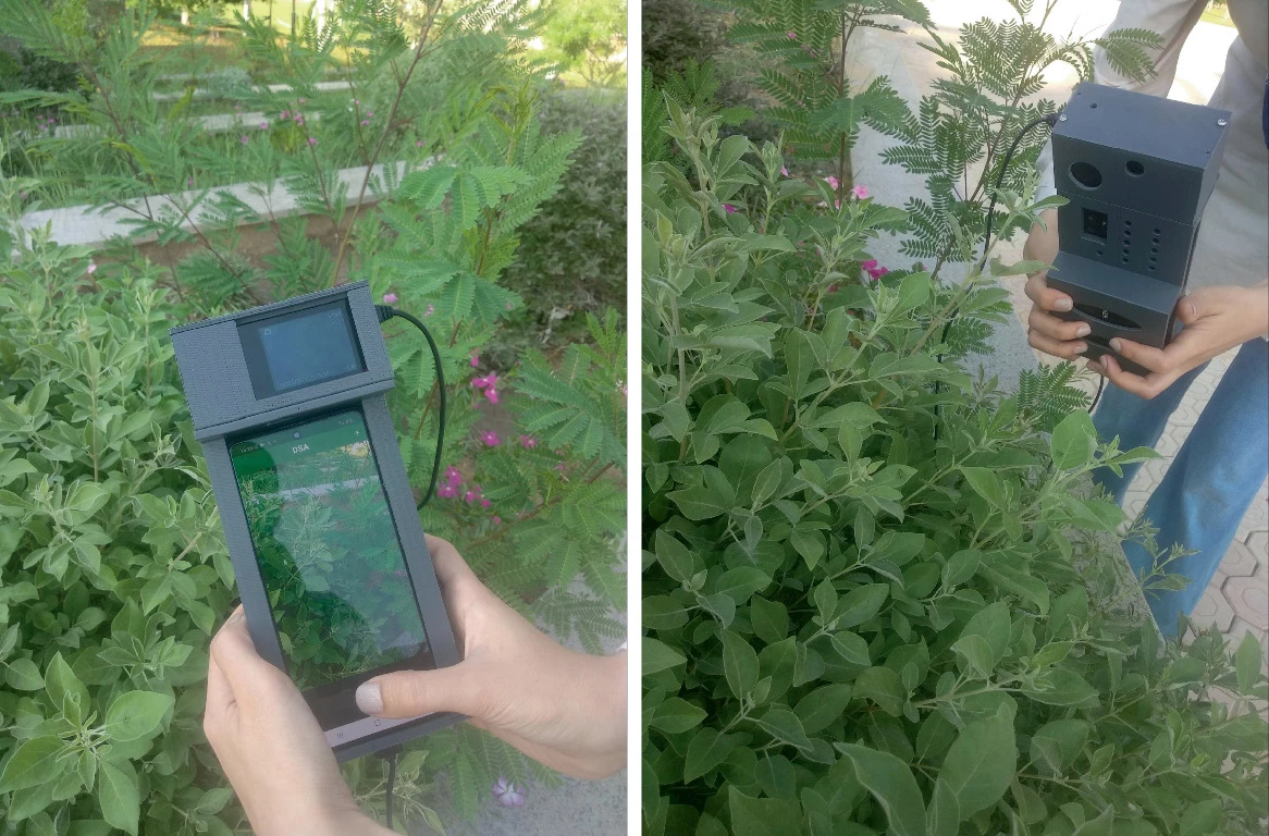
Rice is considered a strategic crop in Egypt as it is regularly consumed in the Egyptian people’s diet. Even though Egypt is the highest rice producer in Africa with a share of 6 million tons per year [5], it still imports rice to satisfy its local needs due to production loss, especially due to rice disease. Rice blast disease is responsible for 30% loss in rice production worldwide [9]. Therefore, it is crucial to target limiting yield damage by detecting rice crops diseases in its early stages. This paper introduces a public multispectral and RGB images dataset and a deep learning pipeline
Automatic Detection of Alzheimer Disease from 3D MRI Images using Deep CNNs
Alzheimer's disease (AD), also referred to simply as Alzheimer's, is a chronic neurodegenerative disease that usually starts slowly and worsens over time. It is the cause of 60% to 70% of cases of dementia. In 2015, there were approximately 29.8 million people worldwide with AD. It most often begins in people over 65 years of age as it affects about 6% of people 65 years and older, although 4% to 5% of cases are early-onset Alzheimer's which begin before this. In 2015, researchers have figured out that dementia resulted in about 1.9 million deaths. Continuous efforts are made to cure the

Dynamic Modeling and Identification of the COVID-19 Stochastic Dispersion
In this work, the stochastic dispersion of novel coronavirus disease 2019 (COVID-19) at the borders between France and Italy has been considered using a multi-input multi-output stochastic model. The physical effects of wind, temperature and altitude have been investigated as these factors and physical relationships are stochastic in nature. Stochastic terms have also been included to take into account the turbulence effect, and the r and om nature of the above physical parameters considered. Then, a method is proposed to identify the developed model's order and parameters. The actual data has

A Framework for Reporting Ergonomic Sitting Posture Anomalies
Application of ergonomics' rules has become a necessity in today's world. Due to the lack of knowledge of what these rules are and the resources needed to fund them, a lot of people develop health issues. One of the most common health issues relate to sitting in a wrong posture for extended period.In this document, a framework that can help in minimizing the existence of the sitting posture anomaly is proposed. This framework takes into account using limited resources as well as being able to apply it in a home environment. © 2022 IEEE.

Using X-ray Image Processing Techniques to Improve Pneumonia Diagnosis based on Machine Learning Algorithms
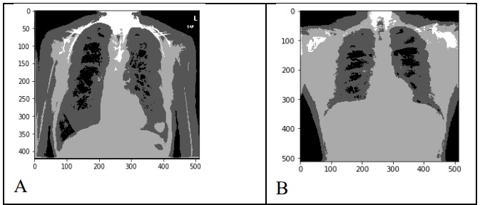
the diagnosis of chest disease depends in most cases on the complex grouping of clinical data and images. According to this complexity, the debate is increased between researchers and doctors about the efficient and accurate method for chest disease prediction. The purpose of this research is to enhance the first handling of the patient data to get a prior diagnosis of the disease. The main problem in such diagnosis is the quality and quantity of the images.In this paper such problem is solved by utilizing some methods of preprocessing such as augmentation and segmentation. In addition are

Edge Detail Preservation Technique for Enhancing Speckle Reduction Filtering Performance in Medical Ultrasound Imaging
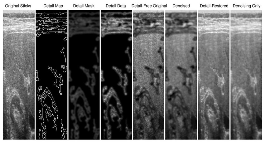
—Ultrasound imaging is a unique medical imaging modality due to its clinical versatility, manageable biological effects, and low cost. However, a significant limitation of ultrasound imaging is the noisy appearance of its images due to speckle noise, which reduces image quality and hence makes diagnosis more challenging. Consequently, this problem received interest from many research groups and many methods have been proposed for speckle suppression using various filtering techniques. The common problem with such methods is that they tend to distort the edge detail content within the image and
Ibn Sina: A patient privacy-preserving authentication protocol in medical internet of things
The prosperous advancement in Medical Internet of Things (MIoT) technologies has hastened the development of healthcare systems. MIoT improves the traditional medical facilities through periodically monitor of patient's health records. Electronic Medical Records (EMRs) are sensitive private data and needs efficient secure and private schemes that interchange these EMRs between healthcare providers and patients. Most of the current privacy preserving schemes do not provide the desired privacy level and suffer from computation and communication overheads. The length of an IDentity-based Strong

Automated multi-class skin cancer classification through concatenated deep learning models
Skin cancer is the most annoying type of cancer diagnosis according to its fast spread to various body areas, so it was necessary to establish computer-assisted diagnostic support systems. State-of-the-art classifiers based on convolutional neural networks (CNNs) are used to classify images of skin cancer. This paper tries to get the most accurate model to classify and detect skin cancer types from seven different classes using deep learning techniques; ResNet-50, VGG-16, and the merged model of these two techniques through the concatenate function. The performance of the proposed model was