
Segmentation of Diabetic Macular Edema in fluorescein angiograms
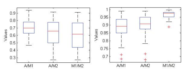
Fundus Fluorescein Angiography (FA) is a powerful tool for imaging and evaluating Diabetic Macular Edema (DME), where the fluorescein dye leaks and accumulates in the diseased areas. Currently, the assessment of FA images is qualitative and suffers from large inter-observer variability. A necessary step towards quantitative assessment of DME is automatic segmentation of fluorescein leakage. In this work, we present an automatic method for segmenting DME areas in FA images. The method is based on modeling the macular image in the early time frame using 2D Gaussian surfaces, which is then subtracted from the late time frame image of the macula to enhance the DME areas. The resulting difference image is then automatically segmented using a Gaussian mixture model classification algorithm. The method was applied to 32 datasets and the results are compared to manual segmentation done by two ophthalmology experts. The results show the potential of the developed method to automatically segment DME. © 2011 IEEE.


