

Segmentation of strain-encoded magnetic resonance images using graph-cuts
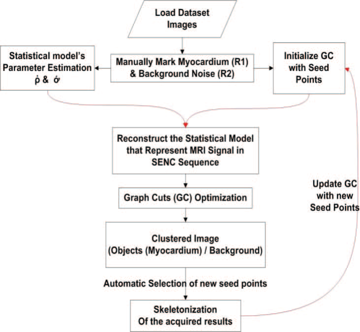
Imaging of the heart anatomy and function using Strain Encoded (SENC) magnetic resonance imaging (MRI) is a powerful tool for diagnosing a number of heart diseases. Despite excellent sensitivity to tissue deformation, the technique inherently suffers from elevated noise level which hinders proper automatic segmentation using conventional techniques. In this work, we propose a method to accurately segment the left ventricle myocardium from strain encoded-MR short axis images. The method is based on a modified formulation of the graph cuts algorithm. A novel cost function based on a probabilistic model for blood and tissue signals is used to achieve proper segmentation results. The method is tested on datasets for eleven human subjects (5 normal and 6 patients). Quantitative evaluation of the proposed method is compared against manual segmentation and the native graph cut algorithm. The results show that the adopted probabilistic model significantly improves the segmentation accuracy compared to the typical cost function of the native graph cuts algorithm. A True Positive and True Negative rates of 92% and 95% respectively have been achieved using the proposed method. © 2011 IEEE.
Related Publications

Hands-on analysis of using large language models for the auto evaluation of programming assignments