
Improved estimation of the cardiac global function using combined long and short axis MRI images of the heart
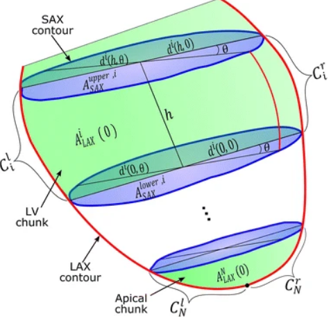
Background: Estimating the left ventricular (LV) volumes at the different cardiac phases is necessary for evaluating the cardiac global function. In cardiac magnetic resonance imaging, accurate estimation of the LV volumes requires the processing a relatively large number of parallel short-axis cross-sectional images of the LV (typically from 9 to 12). Nevertheless, it is inevitable sometimes to estimate the volume from a small number of cross-sectional images, which can lead to a significant reduction of the volume estimation accuracy. This usually encountered when a number of cross-sectional images are excluded from analysis due to patient motion artifacts. In some other cases, the number of image acquisitions is reduced to accommodate patients who cannot withstand long scan times or multiple breath-holds. Therefore, it is required to improve the accuracy of estimating the LV volume from a reduced number of acquisitions. Methods: In this work, we propose a method for accurately estimating the LV volume from a small number of images. The method combines short-axis (SAX) and long axis (LAX) cross sectional views of the heart to accurately estimate the LV volumes. In this method, the LV is divided into a set of consecutive chunks and a simple geometric model is then used to calculate the volume of each chunk. Validation and performance evaluation of the proposed method is achieved using real MRI datasets (25 patients) in addition to CT-based phantoms of human hearts. Results: The results show a better performance of the proposed method relative to the other available techniques. It is shown that, at the same number of cross-sectional images, the volume calculation error is significantly lower than that of current methods. In addition, the experiments show that the results of the proposed model are reproducible despite variable orientations of the imaged cross-sections. Conclusion: A new method for calculating the LV volume from a set of SAX and LAX MR images has been developed. The proposed method is based on fusing the SAX and LAX segmented contours to accurately estimate the LV volume from a small number of images. The method was tested using simulated and real MRI datasets and the results showed improved accuracy of estimating the LV volume from small number of images. © 2016 El-Rewaidy and Fahmy.
Related Publications
Hands-on analysis of using large language models for the auto evaluation of programming assignments