

Transfer Learning in Segmenting Myocardium Perfusion Images
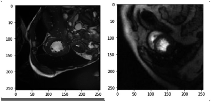
Cardiac magnetic resonance perfusion (CMRP) images are used to assess the local function and permeability of the heart muscle. The perfusion analysis requires the segmentation of cardiac inner and outer walls of the left ventricle (LV). However, the available perfusion datasets are limited or have no annotations. A fair dataset was annotated to employ the latest and most effective Deep Learning (DL) methodologies. In this paper, we employ similar cardiac imaging protocols in terms of cardiac geometry by initially training using CINE images and performing domain adaptation to CMRP images using Unet architectures with different backbones (VGG16, ResNet50, and ResNet152). We also experimented transfer learning (TL) with ImageNet weights by using these architectures separately. Surprisingly, the results were considerable; using CINE images’ weights with ResNet50 as a backbone encoder had better results than other models. This experiment's validation results of dice coefficient, recall, precision, and dice loss results are 0.85, 0.776, 0.94, and 0.148, respectively. The complexity of the data was investigated by finding the intra-observer error using the dice coefficient; the mean result of the intra-observer was 0.796. This study showed how valuable finding TL may help in studying the CMRP images and if the insufficiency of data could be overcome to develop more models to assist in diagnosing heart diseases. © The Author(s), under exclusive license to Springer Nature Switzerland AG 2024.
Related Publications
Hands-on analysis of using large language models for the auto evaluation of programming assignments