

Healthcare
Computer-aided analysis of fluorescein angiograms using colour leakage maps
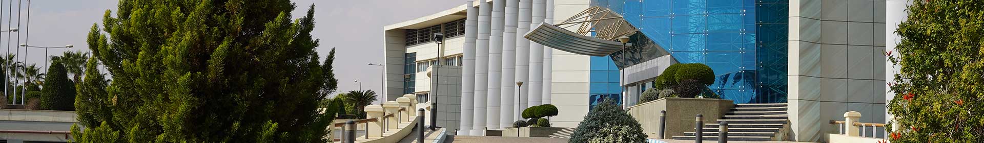
Fundus fluorescein angiography (FFA) is a standard screening and diagnosis technique for several retinal diseases. The analysis of FFA images is performed qualitatively by skilled observers, and thus is vulnerable to inter- and intra-observer variability. In this study, the authors present a method for computer-aided analysis of FFA images. The method is based on generating quantitative colour fluorescein leakage maps (FLM) that mimic the thickness maps generated by the optical coherence tomography (OCT). Results from 64 patients show strong correlation between the FLM and OCT thickness maps
A Survey of COVID-19 Contact Tracing Apps
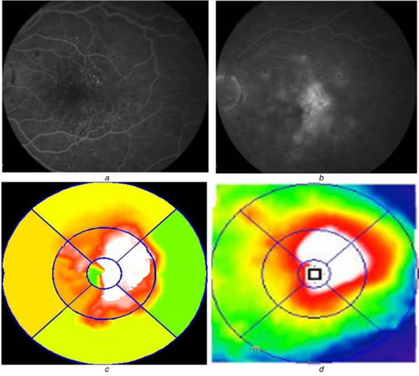
The recent outbreak of COVID-19 has taken the world by surprise, forcing lockdowns and straining public health care systems. COVID-19 is known to be a highly infectious virus, and infected individuals do not initially exhibit symptoms, while some remain asymptomatic. Thus, a non-negligible fraction of the population can, at any given time, be a hidden source of transmissions. In response, many governments have shown great interest in smartphone contact tracing apps that help automate the difficult task of tracing all recent contacts of newly identified infected individuals. However, tracing
A robust quasi-quantum walks-based steganography protocol for secure transmission of images on cloud-based E-healthcare platforms
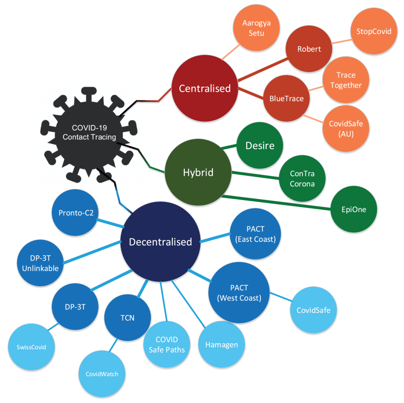
Traditionally, tamper-proof steganography involves using efficient protocols to encrypt the stego cover image and/or hidden message prior to embedding it into the carrier object. However, as the inevitable transition to the quantum computing paradigm beckons, its immense computing power will be exploited to violate even the best non-quantum, i.e., classical, stego protocol. On its part, quantum walks can be tailored to utilise their astounding ‘quantumness’ to propagate nonlinear chaotic behaviours as well as its sufficient sensitivity to alterations in primary key parameters both important
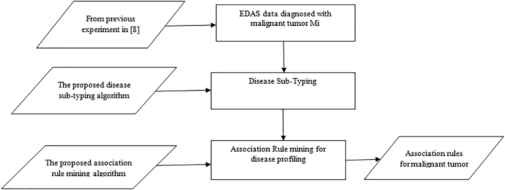
A Theoretical Approach for Correlating Proteins to Malignant Diseases
Malignant Tumors are developed over several years due to unknown biological factors. These biological factors induce changes in the body and consequently, they lead to Malignant Tumors. Some habits and behaviors initiate these biological factors. In effect, the immune system cannot recognize a Malignant Tumor as foreign tissue. In order to discover a fascinating pattern of these habits, behaviors, and diseases and to make effective decisions, different machine learning techniques should be used. This research attempts to find the association between normal proteins (environmental factors) and
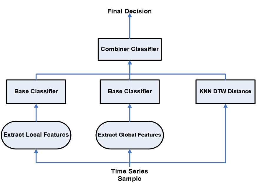
A time series classification approach for motion analysis using ensembles in Ubiquitous healthcare systems
Human motion analysis is a vital research area for healthcare systems. The increasing need for automated activity analysis inspired the design of low cost wireless sensors that can capture information under free living conditions. Body and Visual Sensor Networks can easily record human behavior within a home environment. In this paper we propose a multiple classifier system that uses time series data for human motion analysis. The proposed approach adaptively integrates feature extraction and distance based techniques for classifying impaired and normal walking gaits. Information from body
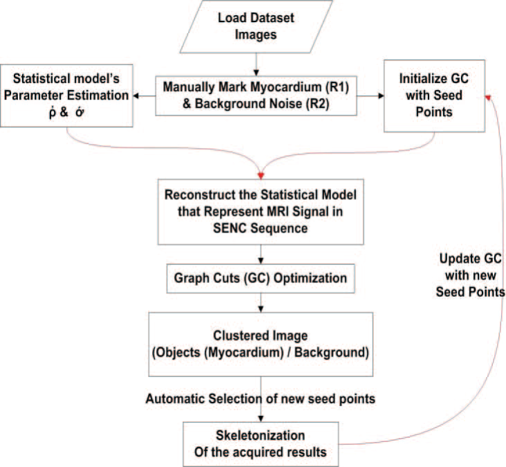
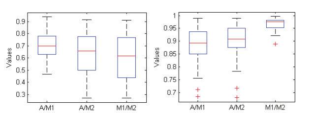
Segmentation of strain-encoded magnetic resonance images using graph-cuts
Imaging of the heart anatomy and function using Strain Encoded (SENC) magnetic resonance imaging (MRI) is a powerful tool for diagnosing a number of heart diseases. Despite excellent sensitivity to tissue deformation, the technique inherently suffers from elevated noise level which hinders proper automatic segmentation using conventional techniques. In this work, we propose a method to accurately segment the left ventricle myocardium from strain encoded-MR short axis images. The method is based on a modified formulation of the graph cuts algorithm. A novel cost function based on a
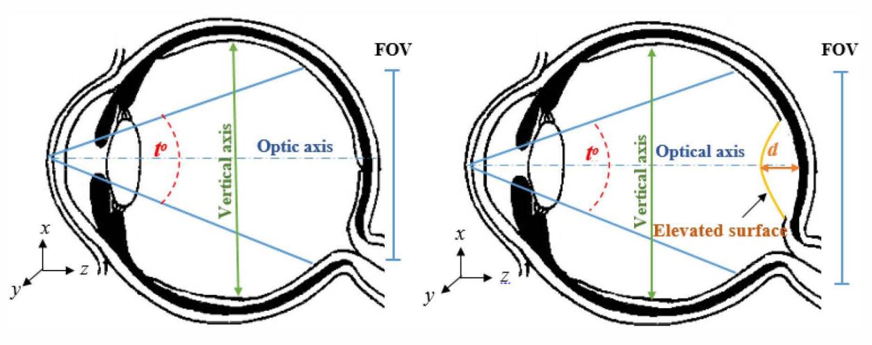
Error analysis of fundus image registration using quadratic model transfformation
Based registration of retinal images proved to be very successful especially for minimally overlapping images. The most commonly used transformation method uses a quadratic model to represent the geometry of the retinal surface. Although this model has been used for more than one decade, there is no literature that studies the model errors for abnormal eye geometries. In this work, we present a study of the registration errors of the quadratic model in case of diseased eyes. The study includes two basic models of the retinal surface for eyes suffering from: myopia; and retinal diseases (e.g
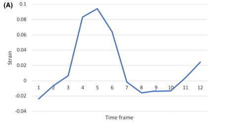
Detecting liver fibrosis using a machine learning-based approach to the quantification of the heart-induced deformation in tagged MR images
Liver disease causes millions of deaths per year worldwide, and approximately half of these cases are due to cirrhosis, which is an advanced stage of liver fibrosis that can be accompanied by liver failure and portal hypertension. Early detection of liver fibrosis helps in improving its treatment and prevents its progression to cirrhosis. In this work, we present a novel noninvasive method to detect liver fibrosis from tagged MRI images using a machine learning-based approach. Specifically, coronal and sagittal tagged MRI imaging are analyzed separately to capture cardiac-induced deformation
Segmentation of Diabetic Macular Edema in fluorescein angiograms
Fundus Fluorescein Angiography (FA) is a powerful tool for imaging and evaluating Diabetic Macular Edema (DME), where the fluorescein dye leaks and accumulates in the diseased areas. Currently, the assessment of FA images is qualitative and suffers from large inter-observer variability. A necessary step towards quantitative assessment of DME is automatic segmentation of fluorescein leakage. In this work, we present an automatic method for segmenting DME areas in FA images. The method is based on modeling the macular image in the early time frame using 2D Gaussian surfaces, which is then
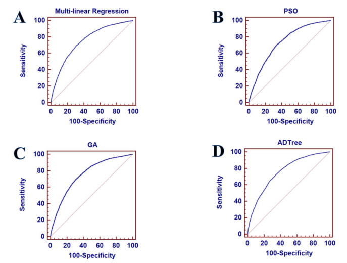
Comparison of Machine Learning Approaches for Prediction of Advanced Liver Fibrosis in Chronic Hepatitis C Patients
Background/Aim: Using machine learning approaches as non-invasive methods have been used recently as an alternative method in staging chronic liver diseases for avoiding the drawbacks of biopsy. This study aims to evaluate different machine learning techniques in prediction of advanced fibrosis by combining the serum bio-markers and clinical information to develop the classification models. Methods: A prospective cohort of 39,567 patients with chronic hepatitis C was divided into two sets - one categorized as mild to moderate fibrosis (F0-F2), and the other categorized as advanced fibrosis (F3