
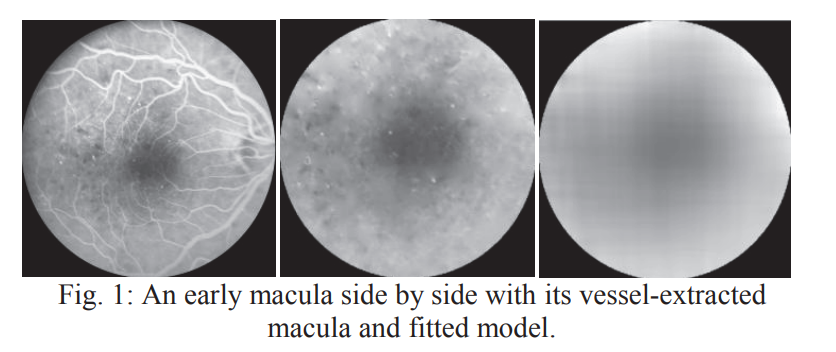
Quantitative assessment of Diabetic Macular Edema using fluorescein leakage maps
Diagnosis of Diabetic Macular Edema (DME) from Fundus Fluorescein Angiography (FFA) image sequences is a standard clinical practice. Nevertheless, current methods depend on subjective evaluation of the amount of fluorescein leakage in the images which lack reproducibility and require well-trained grader. In this work, we present a method for processing FFA images to generate a fluorescein leakage map that can be used for quantitative evaluation of DME. An essential step in the proposed method is to model the spatial distribution of the image intensity within the macula. This model, which represents the non-leaking structures, is then subtracted from the late timeframe image to enhance the areas of fluorescein leakage. The resulting difference image is then mapped with empirical linear transformation to produce a color Fluorescein Leakage Map (FLM) that can be used for quantitative assessment and detection of DME. The method was applied to 13 image sequences for 13 different patients. The resulting FLM maps were found to be correlated with the thickness maps produced by Optical Coherence Tomography (OCT). The relatively high correlation between the FLM and OCT maps show the potential and of using the developed method for quantitatively assess the DME in FFA image sequences. © 2012 IEEE.
Related Publications
Hands-on analysis of using large language models for the auto evaluation of programming assignments