

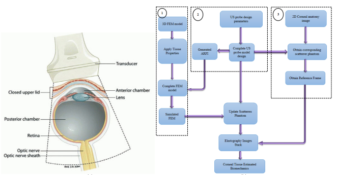
Corneal Biomechanics Assessment Using High Frequency Ultrasound B-Mode Imaging
Assessment of corneal biomechanics for pre- and post-refractive surgery is of great clinical importance. Corneal biomechanics affect vision quality of human eye. Many factors affect corneal biomechanics such as, age, corneal diseases and corneal refractive surgery. There is a need for non-invasive in-vivo measurement of corneal biomechanics due to corneal shape preserving as opposed to ex-vivo measurements that destructs corneal tissue. In this study, a new approach for assessing corneal biomechanics in-vivo non-invasively using ultrasound estimation method with 100 KHz frame rate is proposed. Three models in conjunction with each other are used to study the biomechanics behavior of corneal tissue pre- and post-refractive surgery. These three models are cornea FEM, cornea scatterrer model and ultrasound transducer model respectively. Nine different elastic moduli corneal models are constructed to achieve this goal; 140KPa, 300KPa, 600KPa and 800KPa as post-refractive surgery models and 1MPa, 1.5MPa, 2MPa, 2.5MPa and 3MPa as pre-refractive surgery models respectively. Time-To-Peak (TTP) deformation, deformation amplitude (DA), deformation amplitude ratio at 2 mm (DA ratio 2 mm) and shear wave speed (SWS) are estimated for each of the nine involved corneal models in this study. Results show that TTP is decreasing (6.9 msec. at 140KPa to 5.3 msec. at 3MPa) while increasing the elastic modulus of corneal tissue. Also, DA is decreasing (2 mm at 140KPa to 0.5 mm at 3MPa), while increasing the elastic modulus as well. However, DA ratio shows decrease while increasing of elastic modulus to reflect the difficulty of corneal tissue to deform uniformly at higher elastic moduli. Estimated SWS shows an average accuracy of 98% of the theoretical SWS. © 2013 IEEE.
Related Publications
Hands-on analysis of using large language models for the auto evaluation of programming assignments